Macular Edema
Care Options and Resources for Macular Edema
Macular Edema can make reading, driving, and faces look less clear. It happens when fluid builds up near the retina’s center. Many people notice central vision blurring or dimness first. Some also notice metamorphopsia (wavy lines) on straight edges. Causes vary, so records and eye imaging details matter. This category page helps patients and caregivers compare common causes, testing terms, and care pathways.
Why it matters: Similar vision changes can come from different retinal problems.
Macular Edema: What You’ll Find
This collection focuses on practical navigation for retinal swelling at the macula. It highlights common pathways clinicians discuss after an eye exam. It also points to related condition collections that often overlap in symptoms. For diabetes-related swelling, browse Diabetic Macula Swelling. For vein blockage causes, see Retinal Vein Occlusion.
Many people also compare look-alike conditions with central vision distortion. If clinicians mention abnormal vessel growth, you can review Neovascular Age-Related Macular Degeneration. If notes say “wet AMD,” see Wet Age-Related Macular Degeneration. The goal is easier browsing when paperwork uses unfamiliar terms.
- Common symptom language, including distortion and reduced contrast
- Cause categories, such as diabetes, inflammation, or vein occlusion
- Testing terms like OCT and dye-based imaging
- High-level treatment discussions, including injections, lasers, and steroids
- Administrative notes about prescriptions, verification, and referrals
Video visits use licensed U.S. clinicians in a HIPAA-secure app.
How to Choose
Start with the context in the eye note. Swelling after surgery often gets labeled “cystoid.” Inflammation-related cases may be linked to uveitis (eye inflammation). Diabetes and blood vessel problems often appear in problem lists. A retina specialist visit may include new imaging, even with recent scans.
Details that help compare resources
- Which eye is affected and how quickly symptoms changed
- Recent cataract surgery, eye trauma, or known inflammation history
- Diabetes status, A1C context, and any kidney disease notes
- Blood pressure and vascular risk factors, when listed in charts
- Prior glaucoma or steroid sensitivity history, if documented
- Imaging terms: OCT (retina thickness scan) and angiography references
- Any mention of “center-involving” versus “non-center” swelling
Questions to bring to a clinician
- Which cause seems most likely based on exam and imaging results
- What follow-up schedule is typical for monitoring changes on scans
- Which treatment options fit the suspected cause and eye history
- What side effects matter most for steroid-based approaches
- When to seek urgent evaluation for sudden symptom changes
Safety and Use Notes
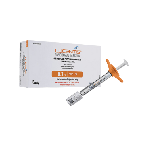
Retinal swelling is a diagnosis that depends on an eye exam and imaging. OCT (optical coherence tomography) helps measure retina thickness and fluid pockets. Fluorescein angiography (dye test) can show leakage patterns in blood vessels. Clinicians use these results to discuss macular edema treatment options. Common categories include anti VEGF injections for macular edema, steroid approaches, and laser photocoagulation macular edema.
- Anti-VEGF (blocks vessel-growth signals) therapy is usually given in-clinic
- Steroid options may include drops, injections, or implants, depending on cause
- Laser can be considered for specific leakage patterns and diagnoses
- Follow-up often relies on repeat imaging to track fluid changes
Clinicians make clinical decisions based on history, exam findings, and imaging.
If vision changes arrive suddenly, the care team may recommend prompt evaluation. Examples include a curtain-like shadow, severe eye pain, or new flashes. Those symptoms can point to problems beyond swelling alone. Medication changes should be reviewed with a licensed clinician. That includes prescription eye drops, diabetes medicines, and blood thinners.
Access and Prescription Requirements
Many prescription items related to retinal care require a valid prescription. Some treatments discussed for this condition happen in an office setting. Others involve home-use medicines for related inflammation or postoperative care. This page also covers practical access notes for Macular Edema management. It focuses on verification, documentation, and safe dispensing standards.
- Prescription-only items require clinician authorization and pharmacy verification
- Names and dates must match the prescription and patient profile
- Some items have handling rules, such as temperature limits or light protection
- Refills and transfers may depend on state rules and prescriber instructions
- Cash-pay options may be available, often without insurance, when permitted
Quick tip: Keep a current medication list saved in the account profile.
When appropriate, clinicians can coordinate prescriptions through partner pharmacies, subject to state regulations.
Related Resources
Many people also want broader vision-support basics, beyond diagnosis terms. For general eye health habits and common misconceptions, see Improve Eyesight Naturally. For a plain-language overview of Macular Edema and common tests, see the American Academy of Ophthalmology overview. For diabetes-related eye disease context, see National Eye Institute diabetic eye disease information.
These resources can make clinic notes easier to interpret. They also help caregivers track which terms map to which tests. When records mention OCT macular edema trends, bring the scan dates. When notes mention fluorescein angiography macular edema patterns, ask what changed. For macular edema vs macular degeneration comparisons, focus on cause and imaging language.
This content is for informational purposes only and is not a substitute for professional medical advice.

Find suitable medication for Macular Edema

Book a telehealth visit to discuss Macular Edema
Find a doctor
Speciality
State

Video Visit
$69

Video Visit
$69

Video Visit
$69

Video Visit
$69

Video Visit
$69

Video Visit
$69

Speciality: Dermatology, Family Medicine, Men's Health, Urgent Care, Women's health
Speaks: English, Spanish, French, Arabic, Portuguese
Video Visit
$69

Video Visit
$69

Video Visit
$69

Video Visit
$69

Video Visit
$69

Video Visit
$69
Frequently Asked Questions
What is macular edema, in plain language?
Macular edema means fluid builds up near the macula, the retina’s center. The macula supports sharp, detailed vision used for reading and faces. Swelling can make straight lines look wavy and reduce contrast. Many causes exist, including diabetes, blood vessel blockages, inflammation, and postoperative changes. A clinician typically confirms it with a dilated eye exam and imaging. The underlying cause often guides which treatment approach is considered.
What symptoms are commonly linked to retinal swelling at the macula?
People often report central vision blurring, dimness, or trouble reading small print. Some notice metamorphopsia (wavy lines), especially on tiles or doorframes. Colors can look less vivid, and contrast may drop. Symptoms can affect one eye or both eyes. These signs overlap with other retinal and optic nerve problems. An eye exam and imaging help clarify what is causing the change. Sudden, severe symptoms may need urgent evaluation.
How do clinicians diagnose macular edema?
Diagnosis usually combines a symptom history with an eye exam and imaging. OCT (optical coherence tomography) is a common scan that measures retina thickness. It can show pockets of fluid and changes over time. Some cases also use fluorescein angiography (a dye-based imaging test) to identify leakage patterns. Clinicians may also look for related findings, like diabetic retinopathy or vein occlusion changes. The testing plan varies by suspected cause and severity.
How is macular edema different from macular degeneration?
They affect the same central retina area but involve different processes. Macular edema refers to swelling from fluid leakage or inflammation. Macular degeneration describes damage and structural changes in the macula over time. Wet (neovascular) age-related macular degeneration can involve leaking abnormal vessels, so symptoms can look similar. Imaging and exam findings help separate these conditions. Charts may also list both when a person has more than one retinal diagnosis.
When are prescriptions required, and how are they verified?
Many prescription eye medicines require an authorized prescription from a licensed clinician. Verification often includes matching the patient name, medication, directions, and prescriber details. Pharmacies also follow state rules for dispensing and refills. Some treatments discussed for retinal conditions happen in-clinic and are not shipped. When a prescription is clinically appropriate, care teams may coordinate options through partner pharmacies. Cash-pay access may be available, often without insurance, depending on the item and location.